BIOMIMESYS® 3D cell culture products
Exploring the Groundbreaking BIOMIMESYS® 3D Cell Culture Products
In the rapidly evolving field of cell culture technology, BIOMIMESYS® has emerged as a cutting-edge solution that is revolutionizing how scientists study human tissues. Unlike traditional 2D cell culture systems, which fail to replicate the complex microenvironments of living organisms, BIOMIMESYS® provides a more accurate and reliable model through its innovative 3D cell culture technology.
Table of Contents
What is BIOMIMESYS®?
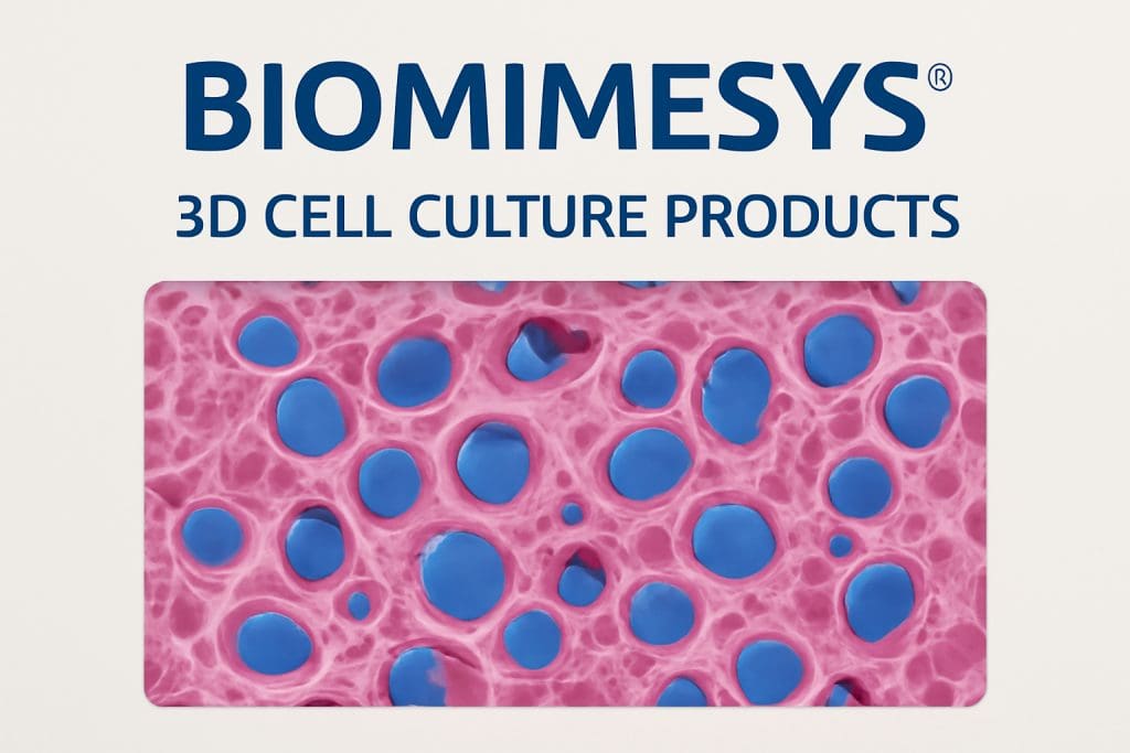
BIOMIMESYS® is a patented 3D cell culture technology that combines the properties of both solid scaffolds and hydrogels. This unique combination enables it to simulate the physiological environment of human tissues, providing a more authentic model for studying cellular behavior, drug testing, and disease modeling. BIOMIMESYS® products recreate the intricate architecture of human extracellular matrix (ECM), which includes various components like collagen, adhesion proteins, and glycosaminoglycans (GAGs), most notably Hyaluronic Acid (HA).
The key to BIOMIMESYS®’s functionality lies in its ability to mimic the natural cell-cell and cell-matrix interactions, which are crucial for understanding the cellular environment in the human body. By offering a 3D environment, BIOMIMESYS® enhances the accuracy of experiments and the potential for translating research into therapeutic applications.
The Hydroscaffold™ Technology
At the core of BIOMIMESYS® is the Hydroscaffold™ technology, a proprietary system that blends the structural properties of a scaffold with the dynamic characteristics of a hydrogel. This allows for the creation of a highly adaptable and customizable matrix that can replicate the cellular microenvironment of various organs or tissues. Depending on the target organ or tissue, the composition and proportion of extracellular matrix components can be adjusted to better mimic the unique characteristics of the tissue being studied.
For instance, the elasticity, porosity, and density of the matrix can be tuned, simulating the diverse conditions found in human organs. The ability to modify the Elastic Modulus (the stiffness of the matrix) and other physical properties of the matrix enables the simulation of tissues ranging from soft, flexible organs like the liver to stiffer tissues such as bone.
Composition of BIOMIMESYS® Matrices
BIOMIMESYS® matrices are primarily made from Hyaluronic Acid (HA), the most abundant GAG in the human body and a crucial component of the ECM. HA is involved in various cellular processes such as cell migration, differentiation, and proliferation, making it an essential part of the matrix for 3D cell culture systems. The matrices also incorporate collagens, which are vital for providing structural support to tissues, and adhesion proteins that facilitate cell attachment and growth.
The innovative manufacturing process of BIOMIMESYS® ensures that the natural properties of HA are preserved, maintaining its essential characteristics for cell growth and development. The combination of HA, collagens, and adhesion proteins creates an ideal environment for cells to behave more like they would in the human body, allowing for better predictability and reproducibility in experiments.
Applications of BIOMIMESYS® 3D Cell Culture
The versatility of BIOMIMESYS® makes it ideal for a wide range of applications in biomedical research and drug development. Some of the key uses include:
- Drug Discovery & Testing: Traditional 2D cultures often fail to predict how drugs will interact with tissues in vivo. BIOMIMESYS® offers a more accurate representation of human tissues, allowing for better screening of pharmaceutical compounds, toxicity testing, and optimization of drug formulations.
- Disease Modeling: By mimicking the cellular microenvironment of specific tissues or organs, BIOMIMESYS® enables researchers to model various diseases more effectively. This includes cancer, cardiovascular diseases, and metabolic disorders, where understanding cell behavior in a realistic 3D environment is crucial for developing targeted treatments.
- Tissue Engineering: BIOMIMESYS® plays a key role in tissue engineering, where it can be used to grow functional tissues for transplantation or regenerative medicine. The ability to tune the matrix composition and structure makes it possible to create tissues with properties similar to those of human organs, offering potential solutions for organ replacement.
- Cell Therapy: In stem cell research and regenerative medicine, creating the right environment for stem cell growth and differentiation is essential. BIOMIMESYS® provides the necessary support for stem cells to thrive and differentiate into specific cell types, facilitating advancements in cell therapy.
Benefits of BIOMIMESYS® Over Traditional Models
- Improved Relevance to Human Biology: Traditional 2D cultures fail to capture the complexity of human tissues. BIOMIMESYS® overcomes this limitation by providing a more accurate representation of the human microenvironment.
- Customization: BIOMIMESYS® matrices can be tailored to replicate different tissues, allowing researchers to focus on specific organs, such as the liver, brain, heart, or skin, based on the needs of their studies.
- Increased Reproducibility and Predictability: Because BIOMIMESYS® better mimics the human body, experiments conducted using this system tend to be more reproducible and yield results that are more predictive of real-world outcomes.
BIOMIMESYS® is a pioneering 3D cell culture technology that is transforming how we study human tissues. By combining the strengths of hydrogels and scaffolds, this technology offers researchers a more accurate, reproducible, and customizable platform for studying cellular behavior, testing drugs, and developing new therapies. With its ability to mimic the extracellular matrix of various organs and tissues, BIOMIMESYS® is poised to become a cornerstone of biomedical research and regenerative medicine, ultimately contributing to more effective treatments and therapies in the future.
Whether for drug development, disease modeling, or tissue engineering, BIOMIMESYS® provides a versatile and powerful tool that brings us closer to truly understanding human biology at the cellular level.